
If the eye becomes injured in the field, swift and appropriate action must be taken to prevent its loss. During your assessment you will seek to determine what part of the eye was injured, to what depth, and its severity. Understanding the anatomy of the eye is important for these assessments, as injuries to certain areas should always raise red flags. Considering the MOI, what are the results of the following inspections:
Try to determine if there is a “globe rupture”. A globe rupture is a loss of integrity of the outer membranes of the eye (sclera and cornea). Any full thickness injury to the sclera and/or cornea-be it blunt or penetrating-is considered a globe rupture. This is one of the most serious types of eye injury, and loss of the eye is a major possibility. If found, try not to let the patient move their eye. Signs include; a “peaked”/bulging pupil, a tear shaped pupil, a mass under-or hemorrhage of-the conjunctiva (the thin clear lining that covers the eye and the eyelids), loss of the iris (there will be no “colored” portion of the eye visible and the pupil may be abnormally large, loss of the lens.

2. Determine if the vision has been affected. Ask your patient if there is blurry vision, double vision, loss of depth perception, or a loss of vision all together.
Does the patient retain a normal field of vision? You should be able to see approx. 95 degrees outward (towards your ear) and approximately 60 degrees inward (towards your nose). You should be able to see 60 degrees up and 75 degrees down, from the center. This translates to a horizontal range of 155 degrees and vertical range of 135 degrees. Top test this, you can extend your forefinger in front of the patient, and ask them to track it with just their eyes (not moving their head). Slow move the finger to the left, then right, then up, then down, asking the patient to tell you when they can no longer see the finger in each field of view. Keep your finger at least 18” from their face when performing the exam.
Visual disturbances after a trauma to the eye are a serious red flag and may mean the retina is damaged. Trauma to the retina can result in blindness, and it requires immediate care. Some visual disturbances which are symptoms the retina is damaged or detached are; flashing lights (aka seeing stars ), floaters (specks, lines or cobwebs in your field of vision), and a shadow appearing in your peripheral vision.
3. Try to determine if there is damage to the bony structure around the eye. Palpate very gently and be especially aware of the presence of any crepitus, as this is a major red flag. Should one eye present as appearing larger than the other after damage to this area, a “blowout” fracture may have occurred. This is a major red flag.
4. Inspect the eyelid and the integrity of the integrity of cornea, sclera, & conjunctiva. Take note of any lacerations, blood, inflammation, redness, etc. You may gently pull the eyelids up and down respectively, and ask the patient to look up/down/left/right to inspect the eye and the lids further.
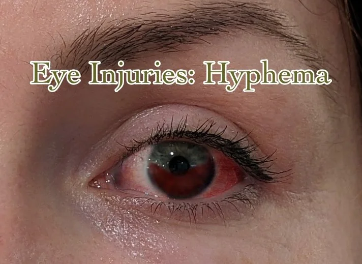
5. Is hyphema (blood in the chambers of the eye) present? This can occur on isolated areas of the eye, or cover the majority. Note the blood will be pooling inside the eye. This can cause very dangerous rises in pressure inside the eye.
6. If no penetrating trauma or globe ruptures are present, can the patient move the eye up, down, left, right? Remember to ask if there is pain or loss of vision when doing so.
7. Does the pupil still respond to light?
Eye injuries of any seriousness must be treated quickly. Should your assessment reveal anything other than mild irritation or lacerations to areas of least concern (the eyelid or outermost lens), then evacuation is highly recommended. Remember, if you must bandage one eye-and it is important for it not to move-then you must bandage both eyes, as they do not track independently.